Mysteries of sleep explored with combined MRI, EEG information
Understanding sleep is no snore to researchers at NIH's National Institute of Neurologic Disorders and Stroke (NINDS) who are studying an intriguing question in sleep science – what changes can we observe in specific regions of the brain in different stages of sleep?
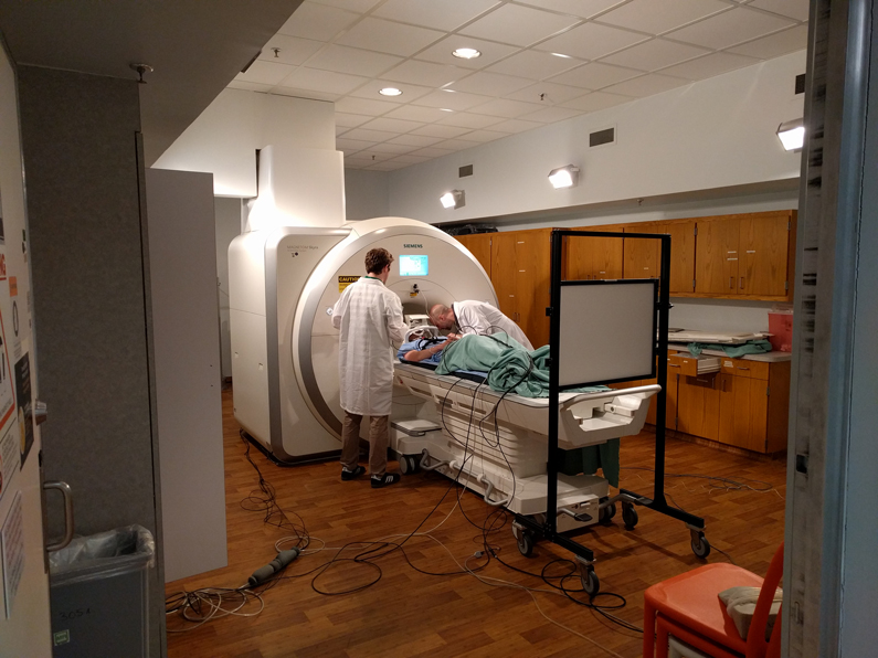
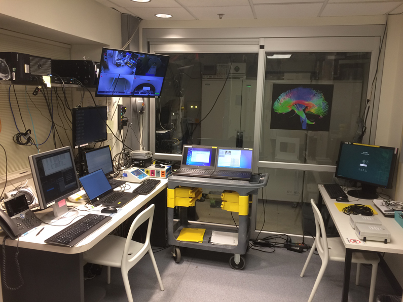
To investigate, a team of researchers is exploring the use of functional magnetic resonance imaging (fMRI) to study brain activity and its changes over time – a different process than the more familiar structural MRI, which simply produces a static image of regions of the brain. fMRI provides three-dimensional, progressive views of activity in the brain in the various stages of sleep.
One of the research team's members, Dante Picchioni, Ph.D., scientist at NINDS said, "We were reviewing the literature, looking for gaps. We saw that very few studies had been conducted using fMRI that measured changes in brain connectivity across time."
Earlier sleep studies relied on electroencephalography (EEG), which measures brain waves in sleep stages, including the familiar rapid-eye-movement stage, commonly known as REM sleep.
"We were looking for a parallel avenue of research that would complement what we had learned using EEG", Picchioni added. "We wanted to see if new research could help further define what sleep actually is and what it does."
The team first completed a pilot study to show that all-night fMRI studies were feasible, and from that, call attention to what kind of expanded study might be possible. According to Picchioni, one surprising finding was the contribution of the autonomic nervous system (a largely unconscious process that regulates bodily activity enclosing heart and respiratory rate, among many functions) and associated non-brain related causes of activity seen in fMRI.
Viewing sleep as a human behavior with four characteristics (no physical motion, a specific posture, immediate reversibility and increasing thresholds for awakening associated with sleep stages), the researchers wanted to correlate fMRI with these observable conditions. By studying new fMRI images, the team is assembling a more complete picture of what the brain activity that makes the progressive sleep stages occur truly looks like while it produces the divergent brain waves associated with different stages of sleep.
Could these fMRI and EEG-aided findings about brain activity in sleep lead to next-phase studies? From what is being learned about the hidden processes in all stages of sleep it might be possible in the future to explore and quantify how duration and progression of sleep stages in specific regions of the brain might affect physical activity, mental insight, acuity and memory while awake the next day.
Picchioni states, "This is an ongoing multi-year study – we're trying to better understand the functions of sleep. Functional MRI, supported by EEG data is helping us in this effort."
- Robert Burleson