Nobel Laureate Discusses Recent Progress in Advanced Microscopy
"One microscope can't do it all, but nothing says you can't have more than one microscope in your box."
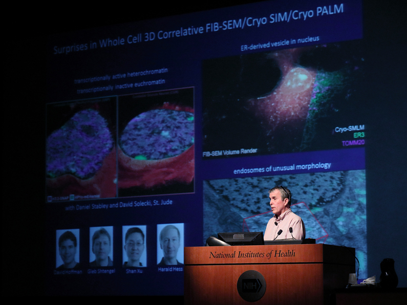
On April 24, Dr. Eric Betzig, of the Howard Hughes Medical institutes' (HHMI) Janelia Research Campus and a 2014 Nobel Laureate for super resolution in microscopy, visited the NIH Clinical Center's Masur Auditorium to address the NIH Director's Wednesday Afternoon Lecture Series to speak about progress in advanced microscopy.
After briefly reviewing the historical development of the microscope and microscopy up through the 1970s, Betzig spoke about significant advances that began in the 1980s and continue to transform microscopy today. He attributes concurrently developing factors - desktop computers used to control microscopes, advances in digital imaging, laser technology and immunofluorescence techniques - which led to a 'Cambrian explosion' of new microscope serving different areas of interest in the biological sciences.
Betzig's own mark on the field came somewhat later when he and longtime friend Dr. Harald Hess joined forces to develop the first super high-resolution, photo activated localization microscopy (PALM) microscope. Betzig and Hess further refined and tested PALM on-site at NIH in collaboration with Dr. Jennifer Lippincott-Schwartz and her team. After moving on to the HHMI Janelia research facility, Betzig and Hess together worked on further experiments with PALM while other investigators also explored its potential. For his pioneering work in developing PALM, Betzig was awarded a Nobel prize in 2008.
Aware of limitations in super-resolution microscopy, Betzig and Hess worked on further refinements including live imaging. Comparing it to seeing the difference of still images of a football game to seeing the game in motion, this PALM variant became known as single-particle tracking (SPT- PALM). Acknowledging the subsequent contributions of numerous other researchers, Betzig explained some of their discoveries and realizations made using SPT-PALM.
His next goal was to develop a microscope for 3-D live imaging. Known as 'lattice light sheet' microscopy, subsequent experiments using this technique led to advances in understanding DNA/RNA transcription and various cellular processes. Drawing inspiration from astronomical research, Betzig's inquiry moved on to the field adaptive optics (AO) and its potential to enhance lattice light sheet microscopy, which he demonstrated with numerous biological examples.
Acknowledging an accessibility problem for labs worldwide to have access to state of the art microscopy, Betzig addressed the challenges to commercial development of advanced and highly specialized microscopes. Because of the enormous interest in his design, the first step, he said, was to thoroughly document and describe the device and subsequently license the design for commercial manufacturing.
With this in process, he was already getting started on the next generation of AO lattice microscope with expanded capabilities. The result of this work was what he terms the "Swiss Army knife" of AO microscopes combining numerous but previously separate operational modes into one compact package at a research-friendly price. The very first of this next generation of microscopes are just starting to be used.
Realizing even as large as the group of potential users may be, the number of technicians needed for constructing and maintaining this new instrument is currently quite small. This can be overcome by commercial development, so Betzig and his team have offered open access to the unit at their faculty at the Janelia Institute, (and soon at University of California/Berkeley) creating strong interest in the immediate scientific community -- and even beyond it, with interest expressed by area tech giants.
Addressing the challenge presented by the enormous volume of data generated from AO experiments, Betzig outlined the interactions between biologists, imaging technicians and data scientists to complete the loop that brings data-driven results back to the biologist, emphasizing the role of the data scientist in this process. As efficiencies are found, the more quickly large datasets can be examined.
Betzig's story is one of determination and relentless experimentation to expand the potential of advanced optical microscopy. He concluded his presentation by stating "…. because of new technologies we have been able to create microscopes to see cellular dynamics with a clarity we could not have imagined even 15 years ago. These are finally allowing us to understand the findings of molecular biology and biochemistry in the context of the dynamic cell. I think that's going to change the way we look at living systems from here on."
- Robert Burleson